If you’ve been scrolling through surgical photo galleries wondering what upper lid blepharoplasty before and after results actually look like in real life, you’re in the right place. This guide breaks down what to expect, how to interpret those photos like a surgeon would, and how healing unfolds over 12 months.
Upper Lid Blepharoplasty Before and After: What You Can Realistically Expect
The transformation from upper lid blepharoplasty is often described as looking like yourself—just more rested and alert. Understanding what typical before and after results look like helps set realistic expectations before your consultation. Here’s what most patients see when comparing their preoperative and postoperative photos:
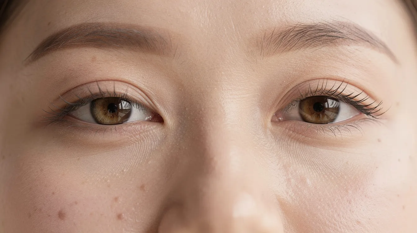
Less hooding over the eyes. The redundant skin that previously folded over the natural eyelid crease or rested on lashes is reduced, creating a cleaner lid surface.
Smoother upper lid contour. Puffiness from herniated fat bags or bulging tissue is flattened, giving a more uniform appearance from brow to lash line.
More visible eyelid crease. The fold that was hidden beneath excess skin becomes apparent, often making eyes appear larger and more defined.
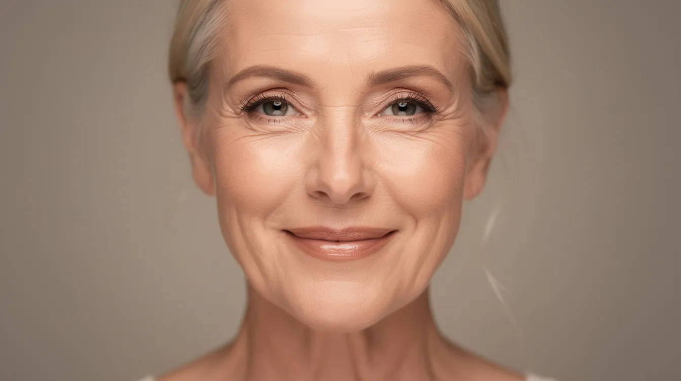
Brighter, more “awake” appearance. The heavy, tired look gives way to eyes that appear refreshed and alert, better matching how patients feel inside.
Most major swelling resolves within 2–3 weeks, allowing patients to return to social settings with sunglasses if needed. However, the “true” after photos displayed in surgeon galleries are typically captured around three months post-surgery, when contour definition becomes clear and residual puffiness has resolved.
The goal of well-performed upper blepharoplasty is a refreshed version of you—not a different eye shape or a pulled, operated appearance. Skilled surgeons preserve the natural architecture while removing only what creates the heaviness.
Scars are strategically placed within the natural upper eyelid crease. In quality before and after galleries, these incision lines are barely visible by 6–12 months, especially with eyes open. When evaluating photos, look for results that appear natural in motion—blinking and smiling should reveal harmony rather than tension or asymmetry.
What Is Upper Lid Blepharoplasty and Who Is It For?
Upper lid blepharoplasty is an outpatient eyelid surgery that removes or reshapes excess upper eyelid skin, underlying orbicularis muscle, and sometimes orbital fat to reduce hooding and restore a more open, youthful appearance. The procedure typically takes 45–90 minutes under local anesthesia with optional sedation.
It’s important to understand the distinction between purely cosmetic upper eyelid surgery and functional surgery performed when excess skin physically obstructs superior vision. Many patients qualify for both designations—their heavy upper eyelids create a tired appearance while also blocking their visual field.
Common indications include:
Hooded upper lids where redundant skin folds over the crease
Excess skin resting on or brushing against lashes
Difficulty applying eyeshadow or eyeliner due to fold interference
Eyes that look tired or aged despite adequate sleep
Documented visual field obstruction for functional cases
Ideal candidates typically share these characteristics:
Adults ranging from 30s to 80s with good general health
Stable vision without uncontrolled dry eye conditions
No active thyroid disease or bleeding disorders
Realistic expectations about outcomes
Non-smokers or those willing to quit before and after surgery
Both men and women of all ethnicities benefit from upper eyelid blepharoplasty. Surgical planning should always respect each patient’s natural anatomy—preserving monolids or epicanthic folds in Asian patients rather than Westernizing features, and maintaining gender-appropriate crease heights (typically 8–10mm in females, 6–8mm in males).
Anatomy Behind Heavy Upper Lids
Understanding basic eyelid anatomy helps patients interpret what’s actually changing in before and after photos and why certain approaches work better for different concerns.
The levator muscle (levator palpebrae superioris) is the primary elevator of the upper eyelid, attaching to the tarsal plate and lifting the lid with each blink. Müller’s muscle provides additional fine-tuned control. Over decades, these structures can stretch—contributing to ptosis, where the lid margin itself droops 1–2mm per decade after age 40.
Levator muscle stretching causes the lid margin to sit lower, making eyes appear smaller and potentially blocking vision—this requires ptosis repair rather than simple skin removal.
Upper lid fat pads (nasal and central compartments) can herniate forward as the orbital septum weakens, creating puffiness or fat bags that many patients notice in before photos.
Skin elasticity loss accelerates with sun exposure and time—collagen decreases roughly 1% yearly after age 30, leading to crepey, redundant folds by the 50s.
A skilled surgeon evaluates all three elements—skin, muscle, and fat—to create a balanced result. Over-resection of any component risks a hollow, over-operated look that appears unnatural in after photos. The best outcomes preserve appropriate volume while removing only what contributes to heaviness.
Why Patients Choose Upper Lid Blepharoplasty: Common Motivations
Patients pursue upper lid blepharoplasty for deeply personal reasons that blend emotional, practical, and functional concerns. Understanding these motivations helps explain why this remains one of the most requested facial procedures in 2026.
Aesthetic rejuvenation. Approximately 70% of patients cite looking older than they feel as a primary driver. Hooded upper eyelids create a tired, aged appearance that doesn’t match internal energy levels. Many patients simply want their eyes to reflect how awake and vibrant they actually feel.
Makeup frustrations. Redundant skin folds make cosmetics application impractical. Eyeshadow disappears into creases within hours, eyeliner smudges against overhanging tissue, and false lashes won’t adhere properly. Women often describe spending years adapting their routine to work around heavy upper eyelids before seeking surgery.
Functional vision concerns. Between 20–30% of patients report measurable visual field obstruction. They describe missing traffic lights in their peripheral vision, needing to tilt their head back to read, or constantly lifting their brows to see clearly. Visual field testing often confirms 20–40% field loss in these cases.
Asymmetry correction. For 15–20% of patients, one upper eyelid sits noticeably lower than the other in before photos. This imbalance draws attention and can make one eye appear perpetually fatigued. Achieving more balanced, symmetric upper eyelids becomes a key surgical goal.
Genetic early intervention. Patients in their 30s–40s with strong family history of heavy upper lids often seek surgery before dramatic drooping occurs. Familial dermatochalasis or ptosis can appear 10–15 years earlier than typical age-related changes.
How to Read Upper Lid Blepharoplasty Before and After Photos
Photo galleries serve as your window into a surgeon’s skill and aesthetic judgment. Learning to evaluate these images critically—the way a surgeon would—helps you identify quality work and set appropriate expectations for your own results.
When reviewing galleries, look for patients whose age, gender, and ethnic background resemble your own. Results in a young woman with minimal skin excess will differ significantly from those in a 70-year-old with decades of sun damage. Comparing like with like gives a more accurate preview of what’s achievable for your anatomy.
Focus on subtle, natural improvement rather than dramatic transformation. Well-performed blepharoplasty reduces heaviness and improves contour without making eyes look “done” or different.
Verify brow position stays stable between before and after photos. If brows appear significantly higher in the “after,” improvements may partly reflect brow elevation rather than eyelid surgery alone. Gallery captions should clearly state when a brow lift was performed.
Assess both open and closed eye views. Eyes should look relaxed and symmetric in both states. Watch for hollowing, over-exposed upper lid platform, or visible asymmetry that suggests over-resection.
Examine consistency across photo pairs. Reputable clinics use identical lighting, camera distance, and neutral expressions for all before and after images. Inconsistent conditions can make results appear better or worse than reality.
Look for results that appear natural in motion. Dynamic photographs or video (increasingly common in 2026) reveal whether eyes move naturally during blinking and smiling, verifying the surgeon prioritizes functional harmony.
Photographic Angles and Details to Notice
Multiple viewing angles provide a more honest, complete picture of surgical results than a single front-facing shot.
Straight-on views assess eye openness, upper lid fold visibility, and side-to-side symmetry. This angle shows whether both eyes appear balanced and alert.
Three-quarter views reveal the transition from eyelid to brow contour and how the upper lid follows the natural orbital rim. This angle catches hollowing or brow changes that front views may hide.
Side-profile views demonstrate how far excess skin previously hung over lashes—often 3–5mm in before photos—versus the cleaner lash line visible after surgery.
Close-up, high-resolution images allow assessment of scar quality, skin texture, and fine lines. These details matter when evaluating whether results appear polished or rushed.
Standardized backgrounds and camera height indicate professional documentation. Clinics serious about transparency use identical setups for all patient photography.
Upper Lid Crease, Brow Position, and Scar Visibility
Crease position, brow shape, and scar quality serve as key markers distinguishing excellent results from average ones.
Natural crease height should match the patient’s gender and ethnicity in after photos—not artificially high or deeply set. Western females typically show creases at 8–10mm from the lash line; males at 6–8mm. Asian blepharoplasty patients may prefer subtler, lower creases respecting natural anatomy.
Brow height consistency between before and after photos verifies improvements stem from the eyelid procedure rather than compensatory brow elevation. Unless a browpexy or endoscopic browplasty is clearly documented, brow position should remain similar.
Scar maturation timeline means photos at 3–12 months post-op typically show a fine, flat line hidden within the lid crease formation—usually invisible with eyes open. Earlier photos may display faint pinkness that continues fading.
Red, raised, or irregular scars are uncommon in expert hands. If gallery photos show visible step-offs or thick incision lines, discuss scar concerns explicitly during consultation.
Final scar refinement continues up to 12 months, so very early after photos may not represent the stable long-term result. Professional galleries specify the post-operative timeframe for each image.
Healing Timeline in Photos: From Surgery Day to Final Results
Understanding the typical 12-month healing trajectory helps you recognize what’s normal at each stage and when you’ll see your true “after” result emerge. Different post-operative periods look distinct in photographs.
First 48–72 hours. Eyelids swell significantly—up to 50% volume increase is normal. Bruising spreads around upper and lower eyelids into a purple discoloration. Cold compresses and head elevation minimize swelling. Most clinics don’t display this waiting period in public galleries, but patient education materials may show this phase.
Days 4–7. Bruising shifts from purple to yellow and green tones as it resolves. Sutures are typically removed around day 5–7. Many patients feel comfortable in public wearing sunglasses during this first week. Swelling remains noticeable but decreasingly tight.
2–3 weeks post-op. Approximately 80% of bruising has resolved. Moderate residual swelling persists—lids may look slightly full in close-up photos but already dramatically improved compared to pre-op heaviness. Most patients return to work during this window.
One month. Early “after” photos become reasonable. Contour improvement is evident, though some firmness or tightness along the crease may persist. Results appear rested and refreshed even if not fully settled.
Three months. This represents the common timeframe for official gallery photos. Swelling has resolved approximately 90%, natural crease definition is apparent, and scars generally appear pale and flat when eyes are closed. This milestone shows reliable contour without lingering puffiness.
6–12 months. Final refinement occurs as tissue fully settles and scars blend seamlessly with the natural crease line. Patients see their stable, long-term appearance. Photos taken at this stage represent the verified final outcome—what verification successful healing looks like.

Case Examples by Age Group
Different decades of life tend to produce distinct before and after patterns, reflecting varying anatomy and surgical goals.
Early 40s patient with genetic hooding. A young man or young woman presenting with hereditary heavy upper eyelids but minimal sun damage or volume loss typically sees striking improvement in crease visibility and makeup space. Skin is resilient, healing is rapid, and scars become nearly invisible by 6 months. These patients often motivated the procedure because family photos revealed they inherited their parent’s hooded appearance decades early.
Mid-50s patient with skin excess and fat protrusion. A 54-year-old patient with combined redundant skin and puffiness from fat herniation demonstrates the skin-muscle-fat technique well. After photos at three months show a softened upper lid contour with better balance between upper and lower eyelids. Volume preservation remains important—removing too much fat creates hollowing that ages the face differently.
Late 60s–70s functional patient. A 69-year-old woman with ptosis blocking her pupil underwent levator advancement plus blepharoplasty with fat repositioning. Her three-month photos highlight both improved superior visual field—less skin draped over the pupil—and natural, conservative reduction of folds. Surgeons working with older patients often preserve more tissue to avoid a gaunt, over-operated appearance on camera and in daily life. These cases may qualify for insurance coverage when documented visual field loss exceeds 30%.
Techniques, Combination Procedures, and Non-Surgical Alternatives
The specific surgical approach influences what you’ll see in before and after galleries. Understanding technique variations helps you recognize why certain results appear more dramatic or subtle than others.
Skin-only upper lid blepharoplasty removes redundant skin without addressing underlying muscle or fat. This approach suits younger patients or those with thin hooding and minimal puffiness. After photos show smoother lid surfaces without significant volume change.
Skin-muscle-fat techniques address more pronounced puffiness common in patients over 50. Surgeons excise conservative amounts of orbicularis muscle and either remove or reposition 1–2mm of orbital fat. After photos demonstrate flatter, non-bulging upper lids while avoiding the hollow, over-resected appearance that signals excessive removal.
Brow lift or browpexy combinations address patients whose heavy appearance stems partly from descended brows. A brow lift can elevate brows 2–4mm when ptosis contributes to upper lid heaviness. Gallery captions should clearly state when combined procedures create the displayed improvement, as brow contour significantly affects apparent eyelid openness.
Upper eyelid ptosis repair (levator advancement or Müller’s muscle procedures) corrects cases where the lid margin itself sits low—not just excess skin covering the lid. When performed alongside bilateral upper blepharoplasty, these combined approaches produce more significant eye opening in after photos. Ptosis repair addresses the muscle weakness while blepharoplasty handles the skin redundancy.
Non-surgical alternatives like botox can create mild lateral brow elevation (1–2mm) lasting 3–4 months—useful for subtle lift without surgery. Fillers may camouflage tear troughs or volume deficits around the eyes. However, neither option removes excess upper eyelid skin. For significant hooding, surgery remains the only solution offering 10–15 year durability versus injectable’s transient effects.
Setting Realistic Expectations and Choosing a Surgeon
Your satisfaction with before and after outcomes depends heavily on clear expectations and thoughtful surgeon selection. Studies show 95% satisfaction when patient expectations align with what surgery delivers, versus 70% satisfaction when mismatches occur.
Seek board-certified oculoplastic or facial plastic surgeons who specialize in periorbital procedures. These specialists typically achieve complication rates below 1% for issues like infection or hematoma. Their galleries should display multiple, high-resolution before and after cases demonstrating consistent quality.
Request an in-person consultation where the surgeon reviews photos of patients with anatomy similar to yours. A thorough evaluation includes discussion of what is—and is not—achievable given your specific skin quality, fat distribution, and structural anatomy.
Articulate your priorities explicitly. Do you want maximum crease show for easier makeup application? Minimal, conservative change? Primarily functional improvement? Clear communication allows the surgeon to create a tailored plan rather than applying a one-size-fits-all approach.
Ask about revision rates and complication management. Reputable surgeons openly discuss their revision rate (typically 2–5%) and how their practice protects patients if adjustments become necessary. Understanding the follow-up process reveals the security service level their practice provides.
Prioritize natural results in galleries. The most impressive transformations aren’t always the most dramatic. Seek surgeons whose before and after photos consistently show natural looking results—refreshed rather than altered, harmonious rather than overdone.
Frequently Asked Questions About Upper Lid Blepharoplasty Before and After
These common questions address specific concerns patients have when evaluating upper lid blepharoplasty galleries and considering the procedure in 2026.
What’s the typical age range for upper lid surgery?
Candidacy depends more on eyelid anatomy and overall health than exact birth year. Patients ranging from early 30s to late 80s successfully undergo upper blepharoplasty. Younger patients often present with genetic hooding, while older patients may have decades of accumulated skin laxity plus functional concerns. Your surgeon evaluates whether your anatomy would benefit from the procedure—not simply your age.
How long do results last?
Most patients enjoy their results for 8–15 years. Aging continues naturally, but from a “reset” baseline visible in your immediate post-operative photos. The skin removed doesn’t regenerate, though remaining tissue continues normal aging. Later photos taken years post-surgery typically show patients still appearing more open and rested than their preoperative state.
When can I return to work and social events?
Most patients feel comfortable resuming normal activities within 7–14 days, depending on bruising patterns and personal comfort with residual swelling. Some return within the first week using sunglasses, while others prefer waiting until most visible signs resolve. Remote work has made early return easier for many patients in 2026.
When can I wear eye makeup again?
Surgeons typically clear patients for gentle eye makeup application around 10–14 days post-op. Avoiding makeup during initial healing protects the incision and reduces infection risk. Once cleared, cosmetics can enhance after photos—and many patients find makeup application dramatically easier without excess skin interfering.
Will insurance cover my procedure?
Insurance may cover functional upper lid blepharoplasty when documented visual field obstruction exceeds specific thresholds—typically 30% field loss or lid-to-pupil distance measuring less than 2mm. Verifies this coverage requires visual field testing and standardized photographs demonstrating the functional impairment. Purely cosmetic procedures remain out-of-pocket expenses. Your surgeon’s office can guide you through documentation requirements.
What about combining upper and lower blepharoplasty?
Many patients address both upper and lower eyelids simultaneously when lower lid bags, wrinkles, or hollows accompany upper lid heaviness. Lower blepharoplasty techniques vary from transconjunctival fat repositioning to skin removal approaches. Combined procedures extend recovery slightly but provide comprehensive rejuvenation in a single surgical session. Combined approaches may also include a chemical peel or laser resurfacing for skin texture improvement.
Conclusion: Interpreting Your Own Potential Before and After
Upper lid blepharoplasty offers natural, long-lasting rejuvenation when approached with realistic expectations and careful surgeon selection. The procedure resets the clock on heavy upper eyelids while preserving your fundamental eye shape and expression—creating improvement that looks refreshed rather than operated.
Use online galleries as a starting point, not a guarantee. Before and after photos demonstrate a surgeon’s capabilities and aesthetic judgment, but your unique anatomy will influence your personal outcome.
Remember that every patient’s “after” is unique. Even when goals are similar, individual skin quality, healing patterns, and technique choices create distinct results. The page of possibilities narrows to your specific journey.
Prepare a list of favorite gallery cases that resemble your own features—similar age, ethnicity, degree of hooding. Bring these examples to your consultation for discussion. This gives your surgeon concrete visual references for understanding your aesthetic goals.
Consider your next step. If upper lid blepharoplasty addresses concerns you’ve been carrying—whether functional vision issues, makeup frustrations, or simply wanting your appearance to match your energy—an in-person evaluation with a qualified specialist clarifies what’s possible for your eyes specifically.
The best results respond to each patient’s anatomy and goals, creating eyes that look naturally brighter without revealing that surgery occurred. That’s the true measure of successful upper lid blepharoplasty before and after.

-2.jpg?width=2000&height=2000&name=Copy%20of%20YouTube%20Thumbnail%20-%20Life-Changing%20Cosmetic%20Surgery!%20(Document)-2.jpg)

