If you've recently had a liposuction procedure or you're preparing for one, understanding how seromas develop and what to do about them can save you weeks of worry. Seromas are a common complication after surgical procedures like liposuction, but they're also highly manageable when caught early.
This guide walks you through how to recognize fluid pockets, what causes seroma formation, your treatment options, and when to contact your surgeon immediately.
Recognizing Fluid Pockets After Liposuction
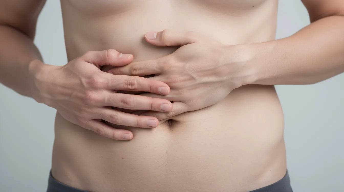
A fluid pocket is a localized collection of serous fluid, primarily lymphatic fluid and plasma, that gathers beneath your skin after surgery. When this fluid collects in the empty space left by fat removal, it forms what's clinically called a seroma. Seromas typically appear as soft lumps under the skin near the treated area.
Common visible signs include:
A soft, fluctuant swelling that feels like a water balloon under the skin
Visible asymmetry compared to untreated areas
Skin that looks distended or stretched over the affected area
Clear or yellowish fluid leakage from the incision site, which may indicate a seroma
Mild tenderness or discomfort without skin discoloration (unless infection develops)
Persistent swelling after surgery may indicate a seroma rather than normal post operative edema.
How to self-check: You can gently press on one side of the swollen area and feel for shifting fluid underneath. This "fluid wave" test helps distinguish fluid filled pockets from firmer tissue swelling. The area will feel jiggly rather than solid. Compare side to side across your body to spot any asymmetry that wasn't there before.
Causes and Seroma Formation
During a liposuction procedure, cannulas break through fat lobules, connective tissue, lymphatic vessels, and small blood vessels. This surgical trauma creates dead space, gaps between tissue layers that the body hasn't yet sealed. The disruption of lymphatic vessels during fat removal means fluid that would normally drain efficiently has nowhere to go.
Seromas result from the body's inflammatory response to the surgical trauma. An inflammatory exudate seeps into these voids, and when your body can't resorb the fluid fast enough, fluid accumulation begins.
Key surgical factors that create dead space:
Large-volume removal of excess fat across multiple body regions
Energy-assisted devices (ultrasound, laser) at high power settings, which increase tissue damage
Wide undermining of skin flaps, especially when liposuction is combined with other procedures
Use of electrocautery, which causes more tissue injury than scalpel techniques
Seromas often form 5 to 10 days after liposuction, frequently appearing right after drain removal. Surgical trauma increases the risk of seroma formation, with chronic cases sometimes persisting for months if left unaddressed.
Risk Factors That Increase Seroma Risk
Not every patient faces the same odds. The incidence of seromas can range from 0.8% to 30% in liposuction cases, depending on technique, patient profile, and procedure scope.
Patient-related risk factors:
Factor | Why It Matters |
|---|---|
Higher BMI (>25) | More dead space, impaired healing; studies show ~1.8% seroma rate vs 0.8% in lower-BMI patients |
Smoking | Delays wound healing, increases inflammation |
Low protein / poor nutrition | Compromises tissue repair |
Diabetes | Impairs blood flow and healing |
Older age | Decreased skin elasticity, reduced lymphatic drainage |
Anticoagulant use | Increases bleeding risk during aspiration |
Procedure-related risk factors:
Removing more than ~100 mL of aspirate per BMI point significantly raises complication rates
Energy-assisted liposuction without carefully controlled device settings
Extensive surgery combining multiple procedures without preventive measures
Absence of drains: one comparative study found seroma in 52% of patients without drains versus only 16% with drains
Engaging in physical activity too soon after surgery can also lead to seromas, as too much movement creates shearing forces between healing tissue layers.
Before your surgical procedure, make sure your surgical team collects a full medical history including BMI, smoking status, nutritional profile, medications, and prior surgeries that may have disrupted lymphatic drainage.
Seroma After Tummy Tuck: Specific Considerations
When comparing tummy tuck risk to liposuction risk, the numbers are striking. In one prospective study of 551 patients, the seroma rate after abdominoplasty was approximately 5.4%, while the liposuction-only group had zero seromas. In massive weight loss patients undergoing a tummy tuck, rates can climb to 30–50%.
The difference comes down to how much tissue is undermined. A tummy tuck involves lifting large skin flaps, creating significantly more dead space than liposuction alone.
Incision sites most prone to fluid buildup:
Lower abdominal flap and infraumbilical region
Pubic area, where fluid can gravitate to the scrotum or labia
Flanks and supraumbilical areas when combining liposuction with abdominoplasty
Surgeons can reduce these risks through techniques like progressive tension sutures and Scarpa's fascia preservation, which maintain lymphatic and venous drainage pathways.
Diagnostic Steps for Fluid Collections
Schedule a clinical exam any time you notice swelling beyond expected post operative edema around day 7 to 10. This is especially important after drain removal or if you experience functional impairment like difficulty walking or stretching.
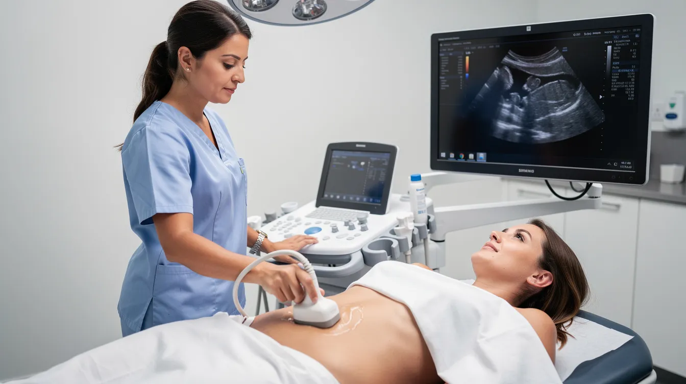
Ultrasound is the imaging method of choice to confirm fluid pockets. It differentiates seroma from hematoma, abscess, or fat necrosis and guides accurate needle placement during aspiration.
Physical Exam Procedures
During a physical examination, your surgeon will document the size and tenderness of any fluid collection using measurements in two dimensions. They'll assess whether the swelling transilluminates (a light shining through clear fluid) and whether it fluctuates when pressed.
Key documentation includes:
Length and width of the swollen area
Whether skin is red, warm, or shows skin discoloration
Comparison with the opposite side of the body
Onset date and changes over time
Imaging And Aspiration Guidance
For deeper or unclear collections, ultrasound-guided aspiration is the most reliable common method. It allows precise needle placement and real-time monitoring of fluid drainage, which is especially important near the surgical site.
If signs of infection are present, such as redness, fever, or cloudy drainage, aspirated fluid should be sent for both aerobic and anaerobic cultures. Not every aspiration requires culture testing, but any turbid or foul-smelling fluid demands it.
Treating Seromas
Treating seromas ranges from watchful waiting to surgical intervention, depending on size and persistence.
Conservative observation: Small seromas may be reabsorbed by the body on their own. Mild seromas that are painless, not under tension, and smaller than 75 mL typically don't require medical treatment beyond monitoring. Most seromas resolve within a month, though some can take up to a year.
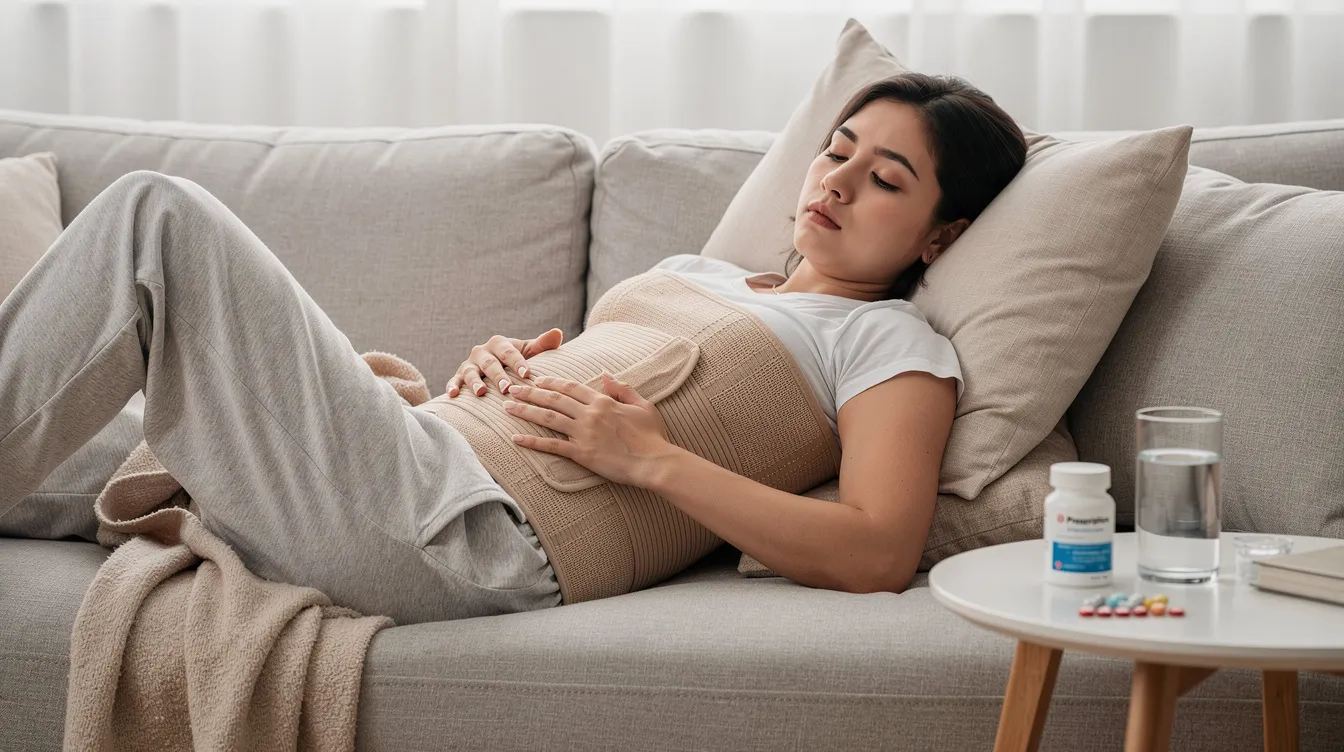
Compression garments: Wearing compression garments as directed helps reduce swelling, minimize fluid accumulation, and promote tissue adherence. They should be snug without restricting blood flow, worn consistently for the first few weeks and often up to 4–6 weeks.
Needle aspiration: For larger seromas exceeding 75 to 100 mL, needle aspiration is typically performed. This involves inserting a small needle or slightly larger gauge needle under sterile conditions, draining as much fluid as possible, and immediately applying compression. Patients may need repeat aspirations every 5–7 days. One to three sessions is typical, though persistent seromas may need more.
Surgical options: When a chronic seroma forms a fibrous pseudocapsule after weeks of recurrence, surgical drainage or capsule excision may be necessary. A surgeon may place a temporary drain, a small tube that channels fluid out of the affected area. In very rare cases, open surgery with debridement is required. These scenarios are uncommon for liposuction-only procedures.
Seromas can become infected or turn into abscesses if left untreated, so don't ignore a fluid collection that keeps returning.
How To Prevent Complications And Reduce Recurrence
Prevention starts before you leave the operating room and continues through your recovery process.
Drain placement: Where appropriate, surgeons place closed suction drains to reduce fluid accumulation during the first few weeks. Keep surgical drains clean to help prevent fluid accumulation and prevent infection at the drain site.
Compression garment adherence: Strict compliance with compression garments is one of the most effective ways to prevent fluid accumulation and reduce fluid buildup. Don't loosen or remove garments prematurely.
Activity restriction: Avoid strenuous activities to reduce seroma risk post-surgery. Follow your post operative instructions precisely. In the first few days, limit movements that cause shearing across treated tissues. Too much movement, bending, and stretching during early recovery directly contributes to seroma development.
Nutrition and lifestyle: A high-protein diet supports tissue repair and helps speed healing. Smoking cessation at least 4–6 weeks before surgery significantly improves wound healing outcomes. Patients with diabetes should have blood sugar well controlled.
When To Seek Medical Attention For Fluid Pockets
Contact your surgeon immediately or seek medical attention if you experience any of the following:
Worsening pain or severe pain at the surgical site that increases rather than improves
Increased swelling that develops rapidly or doesn't respond to compression
Fever, redness, or warmth spreading around the incision site, which are signs of infection
Foul-smelling or purulent drainage, suggesting infected seromas or abscess formation
Cloudy or blood-tinged fluid from the incision, rather than clear fluid
Functional impairment such as inability to move, wound edges separating, or the site starting to leak fluid
Severe symptoms requiring urgent care include increasing swelling and signs of infection. In rare cases, patients on blood thinners may experience bleeding complications during aspiration, so always disclose your full medication list.
Seek medical help if seromas become painful or show signs of infection. Early intervention can prevent complications and further complications like capsule formation.
Post-Treatment Follow-Up And Documentation
Postoperative seromas require structured follow-up to ensure complete resolution and catch recurrence early.
Recommended follow-up schedule:
First visit: ~1 week after treatment or aspiration
During active aspiration: every 5–7 days until fluid collection stops recurring
After resolution: weekly until the surgical team confirms healing is complete
What to document:
Onset date of seroma swelling
Volume of fluid aspirated at each session
Size changes between visits (measured or photographed)
Number of aspirations performed
Any signs of infection or changes in fluid appearance
Monitor for swelling and report changes to your surgeon at every visit. Ultrasound measurements help quantify remaining fluid and confirm that seromas develop no further. Photographs of the area provide a visual record of contour changes across your recovery process.
Most patients find that with consistent compression, restricted physical activity in the early weeks, and close communication with their surgeon's technique and care team, seromas resolve without lasting impact on their results. The key is staying proactive: follow your post operative instructions, attend every scheduled visit, and never hesitate to call if something feels off.
Frequently Asked Questions
What is a seroma after liposuction?
A seroma is a collection of serous fluid, primarily lymphatic fluid and plasma, that accumulates beneath the skin in empty spaces left by fat removal. It typically appears as a soft, water balloon-like lump under the skin near the treated area.
When do seromas typically develop after liposuction?
Seromas often form 5 to 10 days after liposuction, frequently appearing right after drain removal. They result from the body's inflammatory response to surgical trauma and can persist for months if left unaddressed.
What are the main risk factors for developing a seroma?
Risk factors include higher BMI, smoking, poor nutrition, diabetes, older age, anticoagulant use, removal of large fat volumes, energy-assisted devices, extensive combined procedures, and absence of surgical drains. Physical activity too soon after surgery also increases risk.
How can I tell if I have a seroma?
Signs include soft, fluctuant swelling that feels like a water balloon, visible asymmetry compared to untreated areas, stretched skin, clear or yellowish fluid leakage, and mild tenderness. You can perform a fluid wave test by gently pressing and feeling for shifting fluid underneath.
What should I do if I suspect a seroma?
Schedule a clinical exam around day 7 to 10 after surgery, especially after drain removal or if you experience functional impairment. Your surgeon will perform a physical examination and may order ultrasound imaging to confirm diagnosis and guide treatment options.
Learn more: Body Contouring at Leva Medical


-2.jpg?width=2000&height=2000&name=Copy%20of%20YouTube%20Thumbnail%20-%20Life-Changing%20Cosmetic%20Surgery!%20(Document)-2.jpg)


