Your upper eyelid droops, and you're not sure why. Is it a medical condition requiring treatment, or simply the way your eyes are shaped? The answer depends on whether you're dealing with ptosis, hooded eyes, or both. Getting the correct diagnosis matters because these two eyelid conditions look similar but have different causes, risks, and treatment paths.
This guide breaks down the key differences, explains what to look for, and tells you when it's time to see an eye doctor.
Quick Overview: Eyelid Ptosis Versus Hooded Eyes
Eyelid ptosis is a medical condition in which the upper eyelid margin droops abnormally low relative to the pupil, caused by problems with the muscle that lifts the eyelid. Hooded eyes, by contrast, refers to excess eyelid skin that drapes over the eyelid crease and sometimes onto the lash line, without any underlying muscle weakness.
The key distinction between ptosis and hooded eyes is the part that sags. In ptosis, the lid margin itself drops. In hooded eyelids, the skin above the crease folds downward while the margin stays put.
Someone can have both ptosis and hooded eyes simultaneously, which is why a proper diagnosis from an eye doctor is essential when eyelid drooping starts affecting your appearance or your vision.
Why Eyelids Droop: Anatomy, Excess Skin, And Muscle Issues
The levator muscle is the primary muscle responsible for lifting the upper eyelid. It connects to the eyelid through the levator aponeurosis, forming the natural crease. When this muscle or its tendon weakens, stretches, or detaches, the upper eyelid droops and ptosis results.
Dermatochalasis is the medical term for excess skin on the upper eyelid. It develops from a loss of skin elasticity due to aging, sun exposure, and genetic predisposition. Hooded eyes are a normal genetic anatomical variation, and they may become more pronounced with aging as skin laxity increases and orbital fat herniates forward.
Brow descent can also mimic eyelid droop. When the eyebrow drops below the brow bone, it pushes extra skin downward, creating a hooded appearance even when levator muscle function is normal.
Common causes of droopy eyelids include:
Aponeurotic ptosis, which develops from age-related muscle weakening of the levator tendon
Congenital factors present from birth
Nerve damage or nerve disorders such as third cranial nerve palsy or Horner syndrome
Trauma that can damage eyelid muscles, resulting in ptosis
Myasthenia gravis, an autoimmune condition that can cause muscle weakness leading to ptosis
Long-term contact lens wear, which may contribute to aponeurotic ptosis over time
Signs That Indicate A Drooping Eyelid Is Ptosis
The hallmark of true ptosis is a low upper eyelid margin relative to the pupil. Clinicians measure this using MRD-1 (margin reflex distance), which is normally about 3.5 to 5 mm. An MRD-1 of 2 mm or less in primary gaze typically indicates ptosis. Even mild ptosis can produce noticeable asymmetry.
Ptosis can lead to asymmetric-looking eyes and eye strain, especially when one eyelid is more affected than the other. You may catch yourself raising your eyebrows or tilting your head back to compensate.
Watch for eyelid fatigability. If your drooping upper eyelid worsens later in life or later in the day, that intermittent pattern can signal a neuromuscular underlying cause. Ptosis symptoms that fluctuate deserve prompt evaluation.
If you suspect ptosis, try documenting your eyelid position in primary gaze with photographs. Severe ptosis can obstruct the upper field of vision, and ptosis can block the upper visual field or even cover the pupil. Ptosis involves the eyelid margin drooping due to muscle weakness, not just skin overhang.
Signs That Indicate Hooded Eyes From Excess Eyelid Skin
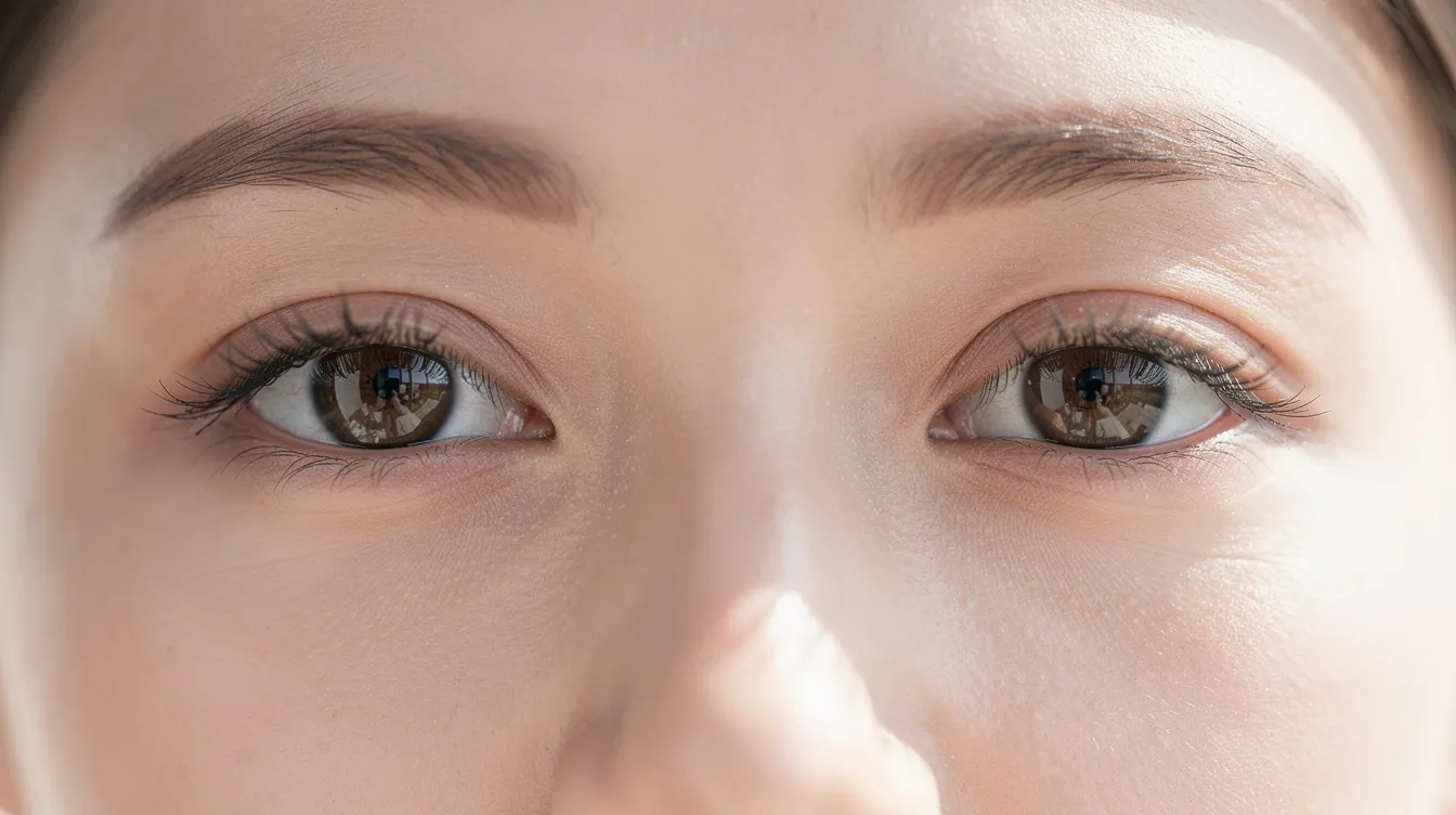
Hooded eyes are caused by excess skin over the eyelid crease. The redundant eyelid skin folds down over the crease and sometimes reaches the lash line, but the eyelid margin itself stays at a normal height. MRD-1 remains within the healthy range.
Hooded eyes are primarily a cosmetic concern, not a medical issue in most cases. Hooded eyes do not affect muscle function or block eyesight, and they rarely obstruct vision in youth. However, as skin laxity progresses with age, severe hooding can eventually reduce the peripheral visual field.
Temporary camouflage options can help you assess the situation. Eyelid tape or adhesive strips physically lift the skin fold. Makeup techniques can create the illusion of a more visible crease. Neither approach addresses a weakened levator muscle, but they can help distinguish a purely cosmetic concern from a functional one.
Congenital Ptosis And Childhood Droopy Eyelid Concerns
Congenital ptosis results from incomplete levator muscle development and is usually present at birth. A child with this condition may have a drooping eyelid in one or both eyes, a poorly defined or absent eyelid crease, and may adopt a chin-up head posture to see underneath the lid.
Ptosis can be congenital or acquired due to aging or trauma. When it appears in infants, the stakes are higher because a lid covering the visual axis can cause deprivation amblyopia, which is permanent vision loss from lack of visual input during critical development.
Early referral to a pediatric eye doctor is essential. If the ptosis is severe enough to threaten vision, surgical intervention may be recommended before age one to two. Even in milder cases, ongoing monitoring of eye movement, refractive error, and alignment is important.
How An Eye Doctor Evaluates Eyelid Function And Ptosis
An examination can determine the difference between ptosis and hooded eyes. The evaluation starts with several key measurements.
MRD-1 quantifies eyelid position. The examiner measures the distance from the corneal light reflex to the upper eyelid margin while your brow is immobilized. A result of 2 mm or less suggests ptosis.
Levator function is tested by measuring how far the lid moves from full downgaze to full upgaze, again with the brow held still. Normal excursion exceeds 12 to 15 mm. Poor function (less than 4 mm) changes which surgical approach is appropriate.
If vision may be obstructed, visual field testing compares your field with eyelids in the resting position versus taped up. Many insurers require documentation showing at least 12 degrees of superior visual field loss. Excess eyelid skin can reduce the peripheral visual field, so this test applies to both ptosis and dermatochalasis.
Clinical photos in multiple gazes, along with all measurements, form the foundation of a treatment plan and any insurance submission.
Non-Surgical Fixes For Droopy Eyelids And Hooded Eyes
Non-surgical options include eyelid tape and Botox for mild cases.
Eyelid tape and adhesive strips provide a temporary mechanical lift. They work for cosmetic reasons or as a diagnostic aid during field testing but offer no lasting correction.
A chemical brow lift using Botox can modestly elevate the brow by relaxing depressor muscles. The effect is limited and cannot address a weakened muscle in the lid itself.
Topical skin-tightening creams and eyelid exercises lack strong clinical evidence. They may feel helpful, but they do not restore levator muscle function or meaningfully reverse skin laxity.
Cold compresses and gentle skin care can reduce puffiness, but they won't correct ptosis or significant hooding.
Eyelid Surgery Options: Blepharoplasty, Ptosis Repair, And Combined Approaches
When drooping eyelids interfere with vision or quality of life, a surgical procedure is often the most effective path.
Upper blepharoplasty is the standard approach to remove excess skin and sometimes fat from the upper eyelid. Upper eyelid blepharoplasty removes excess skin to improve appearance and, when hooding is severe, to restore the visual field. Blepharoplasty is performed under local or general anesthesia depending on the complexity and patient preference. Upper eyelid surgery can improve vision by removing excess skin that blocks the field.
Ptosis repair addresses the actual eyelid function. In a levator advancement, the surgeon tightens or reattaches the levator tendon so the muscle lifts the eyelid to a more normal position. For patients with a severely weakened levator muscle and poor function, a frontalis sling may be used instead. Ptosis surgery tightens or reattaches the levator muscle. Ptosis repair surgery typically takes 1 to 2 hours to complete, and it is typically performed under local anesthesia for adults.
Combined procedures can improve outcomes for patients with both conditions. When sagging eyelids involve both a low margin and excess skin, addressing only one component leaves the other untreated. A ptosis correction paired with blepharoplasty delivers the most complete result.
Before any cosmetic eyelid surgery or functional repair, discuss both expected benefits and surgical risks with your surgeon. Risks include over- or under-correction, dry eye, lagophthalmos, double vision, and the potential need for revision. Serious complications are uncommon but should be understood upfront.

Preparing For Eyelid Surgery And Expected Recovery
Preoperative preparation includes medical clearance, baseline photographs in multiple gazes, and formal eyelid measurements. Your surgeon will review your history for bleeding risk, thyroid disease, dry eye, and neuromuscular conditions like myasthenia gravis.
Recovery typically takes 10 to 14 days after eyelid surgery for most patients. Here's what to expect:
Sutures are usually removed 5 to 7 days post-surgery
Patients may experience swelling and bruising after surgery, peaking around days two through four
Most visible bruising resolves within 2 to 3 weeks
Final results of eyelid surgery refine over several months as tissues settle
During the initial recovery period, avoid heavy lifting, bending, rubbing your eyes, and strenuous exercise. Cold compresses help manage swelling in the first 48 hours. Follow your surgeon's instructions on incision care and sun protection.
Insurance, Medical Necessity, And When Drooping Eyelids Qualify
Not every case of eyelid drooping qualifies for insurance coverage. Hooded eyes that are a purely cosmetic concern typically do not meet criteria. However, insurance may cover ptosis surgery if it obstructs vision.
Criteria that make eyelid surgery medically necessary generally include:
MRD-1 of 2 mm or less in primary gaze
Documented superior visual field loss of 12 degrees or more
Measurable improvement when the lid or skin is taped up
Photographic evidence correlating with test results
Ptosis can obstruct vision and may require medical treatment, so documenting the functional impairment thoroughly is critical. Obtain preauthorization from your insurer before scheduling surgery to avoid denials.
Comparison Checklist: Ptosis Vs Hooded Eyes For Clinical Use
Feature | Ptosis | Hooded Eyes |
|---|---|---|
Lid margin position | Droops below normal; MRD-1 ≤ 2 mm; may cover pupil | Normal height; MRD-1 within 3.5–5 mm range |
Excess eyelid skin | May be present but not primary issue | Primary feature; skin folds over crease and lash line |
Brow position | May compensate by raising; brow ptosis can coexist | May contribute to hooding but lid margin unaffected |
Levator function | Often reduced or impaired | Generally preserved |
Vision obstruction | Likely if lid covers visual axis | Rare unless skin excess is extreme |
Eye shape change | Asymmetry common; eye strain | Symmetric hooding typical; cosmetic concern |
Typical onset | Congenital or acquired ptosis from aging, trauma, nerve damage | Acquired with aging; genetic predisposition |
Primary treatment | Ptosis repair or frontalis sling | Upper blepharoplasty; brow lift if needed |
Both conditions can coexist, complicating treatment options. If you notice a drooping eyelid combined with heavy skin folds, or if your eye shape has changed noticeably, see an eye doctor for a formal evaluation.
When To See A Specialist Or Eye Doctor Immediately
A sudden drooping eyelid that develops over hours or days warrants urgent evaluation. This can signal a stroke, cranial nerve palsy, or other serious medical condition that needs immediate medical treatment.
A child with a droopy eyelid should receive a prompt exam from a pediatric ophthalmologist, especially if the lid appears to block the visual axis. Delays risk permanent vision loss from amblyopia.
For adults with combined features, such as a low eyelid margin, heavy excess eyelid skin, and possible brow descent, consult an oculoplastic surgeon. These specialists are trained to evaluate eyelid function holistically and build a surgical intervention plan that addresses every contributing factor.
Getting the right diagnosis is the first step toward the right treatment plan. If your eyelids are affecting your vision, comfort, or daily life, don't wait. Schedule an evaluation, bring photos taken in natural light, and ask about both ptosis and hooding so nothing gets overlooked.
Frequently Asked Questions
What is the main difference between ptosis and hooded eyes?
Ptosis involves the eyelid margin itself drooping due to muscle weakness. Hooded eyes involve excess skin folding over the eyelid crease while the margin stays at normal height. Both can occur together, requiring proper diagnosis from an eye doctor.
Can hooded eyes affect my vision?
Hooded eyes are primarily cosmetic and rarely obstruct vision in younger people. However, as skin laxity increases with age, severe hooding can eventually reduce the peripheral visual field in some cases.
What causes ptosis?
Ptosis occurs when the levator muscle that lifts the eyelid weakens, stretches, or detaches. Common causes include age-related muscle weakening, nerve disorders, trauma, congenital factors, myasthenia gravis, and long-term contact lens wear.
How do doctors measure whether I have ptosis?
Doctors measure MRD-1, the distance from the corneal light reflex to the upper eyelid margin. Normal range is 3.5 to 5 mm. An MRD-1 of 2 mm or less typically indicates ptosis. Levator muscle function is also tested.
When should I see an eye doctor about droopy eyelids?
Seek evaluation if drooping affects appearance or vision, worsens throughout the day, causes asymmetry, or if eyelid position changes over time. Congenital ptosis in infants requires early referral to prevent vision loss from deprivation amblyopia.
Learn more: Body Contouring at Leva Medical

-2.jpg?width=2000&height=2000&name=Copy%20of%20YouTube%20Thumbnail%20-%20Life-Changing%20Cosmetic%20Surgery!%20(Document)-2.jpg)


