Fibrosis after liposuction is one of the most common postoperative concerns patients face, yet it remains widely misunderstood. Whether you've recently had a liposuction procedure, you're a surgeon refining aftercare protocols, or you're a therapist guiding patients through recovery, understanding how fibrosis develops and how to address it early makes a measurable difference in outcomes.
This guide covers what lipo fibrosis actually is, how to recognize fibrosis symptoms, evidence-based professional treatments, at-home strategies to support healing, and when surgical intervention becomes necessary. Early intervention can prevent severe fibrosis from developing, so the sooner you act, the better your results.
Learn more: Body Contouring at Leva Medical
TL;DR
Liposuction fibrosis is the formation of excess fibrous tissue beneath the skin when the body's normal healing process becomes exaggerated after fat removal.
Common symptoms include hard lumps beneath the skin, uneven skin texture, tightness, localized discomfort, and skin dimpling.
Main professional treatments include manual lymphatic drainage, ultrasound therapy, shockwave therapy, corticosteroid injections, and surgical revision.
Wearing compression garments consistently and starting lymphatic drainage massage early are two of the most impactful steps to reduce swelling and minimize scar tissue formation.
What Is Liposuction Fibrosis?
In simple terms, fibrosis refers to the abnormal or excessive buildup of dense scar tissue in the subcutaneous layer where fat cells were removed during lipo surgery. It develops as the body heals from surgical trauma.
Here's the mechanism: a liposuction procedure creates tissue injury through cannula passes and suction. This triggers inflammation, which activates fibroblasts to deposit collagen and repair the damage. Normally, collagen production is balanced by remodeling over weeks to months. Fibrosis occurs when collagen deposition outpaces resorption, creating hardened scar tissue, bands, or nodules. Collagen buildup, fat necrosis, and chronic inflammation all contribute to this process, especially when disrupted lymphatic circulation allows fluid accumulation to persist.
Fibrosis Symptoms After Liposuction
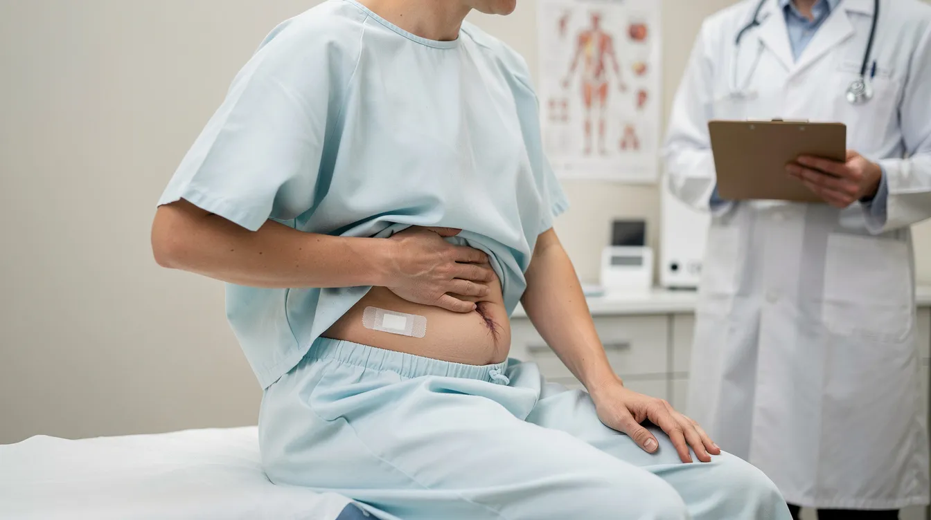
Fibrosis is not the same as normal swelling after liposuction. Here are the palpable signs to watch for:
Hard lumps under the skin, sometimes described as hard lumps beneath the surface
Linear bands or rope-like cords of firm tissue
Skin dimpling, which is a common sign of fibrosis after liposuction
Uneven skin or bumpy surface texture in affected areas
Areas that feel "stuck down" or rigid
Fibrosis feels like tight, firm areas beneath the skin. Symptoms of fibrosis include hard lumps and uneven skin texture, and they often appear 2 to 6 weeks post-surgery. Track your swelling duration carefully. Normal swelling decreases over the first few weeks. If swelling persists beyond 4 to 6 weeks, especially asymmetrically, it may signal developing fibrosis rather than routine recovery.
Report any increased or persistent pain in treated areas to your surgeon, as this may indicate fibrosis. Contour irregularities like dents, ridges, or restricted movement are also warning signs. Persistent fibrosis can disrupt the final aesthetic results post-liposuction.
How Fibrosis Develops: Surgical Technique And Risk Factors
Surgical technique plays a direct role in fibrosis risk. Energy-assisted liposuction (VASER, radiofrequency) increases cumulative tissue trauma and inflammation. Superficial liposuction or aggressive suctioning with large cannulas and repeated passes damages the subdermal plexus and lymphatics. Research shows that high-definition liposuction carries approximately a 2.7% rate of transient soft-tissue fibrosis among minor complications. Aggressive liposuction techniques can lead to higher fibrosis risk, and factors increasing fibrosis risk include aggressive surgical techniques and inadequate compression.
Patient risk factors include:
Higher BMI increases the risk of fibrosis after liposuction
Older patients have a higher risk of fibrosis after liposuction
Smoking increases the risk of developing fibrosis post-liposuction
Previous surgeries in the same area raise fibrosis risk after liposuction
Poor skin elasticity or genetic tendency toward keloid scars
Fibrosis can occur due to poor post-operative care after liposuction
Document every operative detail for follow-up: device type, energy levels, cannula size, number of passes, and any complications such as seroma or hematoma. This information helps guide treatment if fibrosis develops.
Diagnosis And When To See A Professional
If you suspect post liposuction fibrosis, schedule a physical examination with your surgeon. They'll palpate for nodules, bands, and firmness, and visually inspect for contour irregularities.
When diagnosis is unclear, ultrasound imaging and elastography can distinguish fibrotic tissue from fluid collections or residual fat. These tools quantitatively measure fibrosis severity and help localize affected areas.
Seek care immediately if symptoms worsen beyond normal healing timelines, if painful fibrotic areas persist, if skin color changes appear near fibrotic bands, or if you experience limited mobility or functional impairment.
Professional Treatments To Manage Fibrosis
The approach to treat fibrosis follows a stepwise escalation:
Conservative: lymphatic drainage, therapeutic ultrasound, compression garments to soften early scar tissue
Intermediate: shockwave therapy, radiofrequency, laser therapy
Medical injections: intralesional steroids, enzymatic options
Surgical revision: subcision, excision, fat grafting (last resort)
Prioritize early lymphatic therapy for softening healing tissue. Injections or devices become appropriate when conservative measures plateau after several weeks. Surgical revision remains the last-resort option due to the risk of further surgical trauma and more scar tissue.
Patients in New York City have access to advanced recovery resources for managing fibrosis, but regardless of location, seek certified providers experienced in post-surgical rehabilitation.
Lymphatic Drainage Massage
Lymphatic drainage massage should start within days after surgery, as soon as your surgeon clears you. Many protocols recommend beginning within 1 to 2 weeks. Lymphatic drainage massage reduces swelling and fibrosis risk by improving fluid movement and clearing accumulated fluid.
For early-stage fibrosis, aim for 2 to 3 sessions per week over several weeks. A clinical trial with 18 women demonstrated that 12 sessions of combined ultrasound and manual lymphatic drainage produced statistically significant reductions in fibrosis and pain. Always refer to certified therapists trained in lymphatic massage therapy and post-surgical rehabilitation. Improper massage performed too deeply or too soon may worsen inflammation.
Ultrasound And Radiofrequency Therapies
Ultrasound therapy uses sound waves to treat fibrosis non-invasively. Applied once or twice weekly after initial healing, therapeutic ultrasound reduces tissue fibrosis, swelling, and pain, particularly when combined with lymphatic massage.
Radiofrequency stimulates collagen remodeling, softens fibrotic tissues, and improves skin texture. Laser therapy breaks down tough tissues caused by fibrosis and can address contour deformities. A study using a 1,060 nm diode laser on patients at least six months post-surgery showed meaningful contour improvement and smoothing.
Document the device type, energy settings, and session count. Overuse early or at high energy can cause burns or trigger fibrosis rather than resolve it.
Steroid Injections And Enzymatic Options
Corticosteroid injections (such as triamcinolone) are appropriate for dense nodules or existing fibrosis unresponsive to massage and device therapies. Steroid injections target fibrous tissues to decrease inflammation and suppress fibroblast activity.
Treatments are typically spaced weeks apart. Monitor response carefully after each injection. Risks include skin atrophy, pigment changes, and telangiectasia. Enzymatic options like collagenase are emerging but currently lack robust randomized controlled data.
Surgical Technique For Fibrosis Release
Surgical intervention is indicated when non-surgical approaches have failed over 6 or more months, fibrosis is dense and widespread, or contour deformity causes functional discomfort. Procedures include subcision (cutting fibrous bands), excision of fibrotic nodules, fat grafting, or tummy tuck if skin redundancy exists.
A case series of 23 patients undergoing surgical correction achieved near-total resolution of irregularities at 12-month follow-up. Evaluate surgeon experience in revision work carefully and ensure the approach minimizes additional tissue trauma.
Compression Garments To Support Healing
Compression garments support healing and reduce swelling post-surgery. Wear them 24 hours per day for the first 2 to 4 weeks, with many protocols recommending continued wear for 8 to 12 weeks. Wearing compression garments reduces swelling and fibrosis risk by limiting fluid retention and supporting tissue adherence.
Check fit regularly. Garments that are too loose leave pockets for fluid accumulation. Those that are too tight create pressure points and may cause skin irritation. Replace worn garments as they lose elasticity over weeks.
At-Home Care To Reduce Swelling And Support Healing
Start gentle lymphatic massage at home after surgeon clearance. Avoid aggressive deep massage early, as it can worsen inflammation or cause seroma.
Staying hydrated helps flush toxins and supports healing. Aim for adequate daily water intake.
Eat a balanced diet rich in anti inflammatory foods: omega-3 fatty acids, antioxidants, vitamins A, C, and E. Minimize processed sugars that can promote chronic inflammation.
Begin light movement early. Same-day ambulation and gentle range-of-motion exercises improve lymphatic circulation and promote healing without stressing healing tissue.

Lymphatic Massage: Timing, Benefits, And Protocol
Start lymphatic drainage early, ideally within the first 1 to 2 weeks per surgeon guidance. The earliest safe start depends on wound healing, absence of infection, and physician clearance.
Typical session goals include reducing edema, mobilizing fluid, softening tissue, and reducing pain. The São Paulo trial showed that about 37 to 50% of patients still had fibrosis at final assessment, but severity was meaningfully reduced. Track improvements between sessions using consistent grading (visual and palpable scoring, photos, and patient-reported pain). This helps determine whether to escalate to intermediate or medical interventions.
Post Liposuction Recovery Process And Timeline
Timeframe | What to Expect |
|---|---|
Days 1–3 | Peak edema, bruising, tightness |
Weeks 1–2 | Swelling persists, firmness begins; compression critical |
Weeks 3–4 | Bruising subsides; some firmness is part of the normal healing process after liposuction |
Weeks 4–6 | Nodules or bands may become evident if fibrosis is forming |
Weeks 6–12 | Remodeling phase; persistent fibrosis should improve with care |
3–12 months | Maturation; modest improvement continues |
Fibrosis symptoms often appear 2 to 6 weeks post-surgery. Schedule regular postoperative follow-ups at weeks 1, 2, 4, 8, and then at 3 and 6 months. Each visit should assess swelling, skin texture, contour, and compliance with the recovery process.
Preventing Fibrosis: Surgical Technique And Early Care
Choose experienced, board-certified surgeons who minimize tissue trauma through appropriate cannula sizes, controlled energy delivery, and avoidance of excessive passes. The concept of hyperinflammatory fibrosis, identified in 2026 research, emphasizes that cumulative energy delivery in high-definition liposuction is a key risk factor for fibrosis development.
For larger treatment areas, consider staging procedures over multiple sessions to reduce cumulative surgical trauma and prolonged edema. Proper post-operative care can mitigate the effects of fibrosis. Follow post operative care instructions strictly: early compression, ambulatory movement, starting lymphatic drainage early, and avoiding premature strenuous activity. Avoiding smoking improves healing and reduces fibrosis risk. Following post-operative care instructions minimizes complications and is the single most controllable factor in minimizing fibrosis and preventing fibrosis altogether.
Skin Tightening And Aesthetic Corrections After Fibrosis
For addressing fibrosis that has left contour irregularities, noninvasive options include radiofrequency, diode laser, and ultrasound-based skin tightening, typically applied 3 to 6 months after surgery once tissue has stabilized. These improve skin elasticity and smooth surface texture.
Fat grafting corrects contour depressions and volume deficits where fibrosis or tissue loss created asymmetries. For cases involving skin redundancy (particularly the abdomen), a combined approach with abdominoplasty and fibrosis release may be discussed with your surgeon.
When To Consider Liposuction Revision
Objective criteria for revision candidacy include:
Severe persistent fibrosis unresponsive to conservative care beyond 6 to 12 months
Contour deformities, deep depressions, or ridges
Functional impairment or ongoing pain
Patient dissatisfaction after exhausting noninvasive modalities
Wait at least 12 months post-lipo for full maturation of scar tissue and skin contraction before pursuing liposuction recovery revision. Revisions performed too early risk worsening fibrosis with unpredictable results. For complex cases involving energy devices or large treatment areas, obtain second opinions and ensure the revision plan addresses root causes.
FAQs
Can fibrosis resolve on its own? Yes. Mild fibrosis often softens over 2 to 4 months with basic care. Fibrosis is often a temporary healing stage rather than a permanent issue. Moderate cases may take 3 to 6 months with professional treatments. Severe fibrosis benefits from targeted intervention, though full resolution isn't always guaranteed.
How is fibrosis definitively diagnosed? Through clinical physical examination (palpation, visual inspection) combined with imaging like ultrasound or elastography. Distinguishing fibrosis from seroma, hematoma, or residual fat is critical for choosing the right approach to manage fibrosis and cure fibrosis where possible.
When should I contact my surgeon? If firm, tender nodules persist beyond 4 to 6 weeks, if contour irregularities appear, if pain increases rather than decreases, or if you notice skin color changes near fibrotic areas. Early detection allows you to reduce inflammation before the tissue becomes dense and resistant to conservative treatment.
Conclusion And Next Steps
Managing post liposuction fibrosis comes down to three things: recognizing symptoms early, following a structured care protocol, and knowing when to escalate from at-home strategies to physical therapy, device-based treatments, or surgical revision. Most patients see meaningful improvement when they commit to wearing compression garments, maintain a balanced diet, stay hydrated, and start lymphatic drainage early in their recovery process.
If you're noticing hard lumps, uneven skin, or persistent firmness beyond normal healing timelines, don't wait. Schedule a professional evaluation with your surgeon or a certified lymphatic therapist. The window for effective conservative treatment narrows as fibrosis matures, so acting early is the most reliable way to protect your results.
Frequently Asked Questions
What is fibrosis after liposuction?
Fibrosis is excessive buildup of dense scar tissue in the area where fat was removed. It develops when the body's collagen production outpaces resorption during healing, creating hardened tissue, bands, or nodules beneath the skin.
What are the signs of developing fibrosis?
Signs include hard lumps under the skin, linear bands of firm tissue, skin dimpling, uneven texture, and areas that feel rigid or stuck down. These symptoms typically appear 2 to 6 weeks post-surgery and persist beyond normal swelling timelines.
What factors increase the risk of fibrosis?
Risk factors include aggressive surgical techniques, higher BMI, advanced age, smoking, previous surgeries in the same area, poor skin elasticity, and inadequate post-operative compression. Energy-assisted liposuction and superficial liposuction also increase risk.
How is fibrosis diagnosed?
A surgeon performs physical examination, palpating for nodules, bands, and firmness. Ultrasound imaging and elastography can distinguish fibrotic tissue from fluid or residual fat, helping measure severity and localize affected areas.
What are the treatment options for fibrosis?
Treatment escalates progressively: early conservative care includes lymphatic drainage, compression garments, and ultrasound therapy. Intermediate options include shockwave therapy and radiofrequency. Injections and surgical revision are reserved for persistent cases.

-2.jpg?width=2000&height=2000&name=Copy%20of%20YouTube%20Thumbnail%20-%20Life-Changing%20Cosmetic%20Surgery!%20(Document)-2.jpg)


